The fruit fly sheds new light on the genetic control of organ formation

2018-10-10
Based on the study of the formation of legs in the fruit fly (Drosophila melanogaster), researchers at the Autonomous University in Madrid (UAM) and the Severo Ochoa Molecular Biology Centre have managed to describe the relationship between genetic regulation and the cell mechanisms controlling the acquisition of an organ’s form. The results are published in the journal PLoS Genetics.
Researchers at the Autonomous University in Madrid (UAM) and the Severo Ochoa Molecular Biology Centre (a joint UAM-CSIC research centre) have the model organism Drosophila melanogaster, or fruit fly, to clarify how genes control the cell processes determining the shape of a developing organ.
Specifically, the authors of the paper, Sergio Córdoba and Carlos Estella, used the process for the development of leg joints in the fly to try to shed light on the relationship between genetic regulation and shape acquisition, a central issue (albeit little understood) in the field of developmental biology.
For the authors, over and above the particular matter of how leg joints are formed in Drosophila, the relevance of their paper consists in having been able to combine the genetic regulation and morphogenesis processes in a developmental context.
“We have been able to trace the formation of joints from the genetic sub-division of the leg to the cell behaviours that sculpt the shape of the epithelium, and to identify the dysfusion gene as a fundamental element to combine these two processes.”
Genetic origami
The instructions governing the formation of organisms are encoded in the genome. Therefore, for the organism to acquire its shape, size and function, this genetic information must be translated into such cell behaviours as proliferation, differentiation and cellular and tissue movements.
This process for shape acquisition is known as morphogenesis. A very intuitive way to understand the morphogenesis of epithelials (cell layers) is to compare this process to origami, the art of creating figures by folding thin sheets of paper.
In origami, a series of instructions indicate how to fold the paper to obtain the three-dimensional figure. In the same way, genetic information acts during morphogenesis as the instructions indicating the shape the epithelial has to adopt, through the control of the different cell processes (which can be compared to the folds in the paper).
The paper used the formation of fly leg’s joints as a model because there is extensive knowledge about pattern formations in legs. In other words, the genes involved in giving identity to the various segments of the leg and how their expression is regulated are known to a very high degree of detail.
In fact, the group led by Carlos Estella published in 2014, also in PLoS Genetics, a study in which the dysfusion (dysf) gene was identified as the absolutely essential factor for the formation of leg joints.
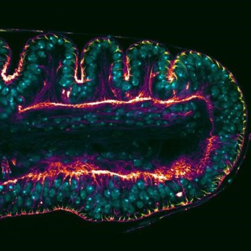
In this new study, the authors go one step further to describe the elements controlled by the dysfusion gene and responsible for directing the changes in the shape of the cells to form the epithelial folds that give rise to the joints.
The study concludes that apical constriction mediated by Rho1 protein, a process widely described for the shape change of epithelial tissues, is controlled by dysf to bring about these characteristic folds in the imaginal disc of a leg.
____________________
Bibliographical reference:
Córdoba S., Estella C. (2018) The transcription factor Dysfusion promotes fold and joint morphogenesis through regulation of Rho1. PLoS Genet. Doi: 10.1371/journal.pgen.1007584

