Actualidad
Premios y Reconocimientos
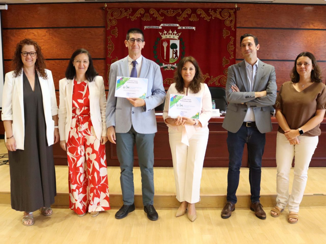
IV Jornada de Tutores Profesionales: un homenaje a quienes enseñan desde la experiencia
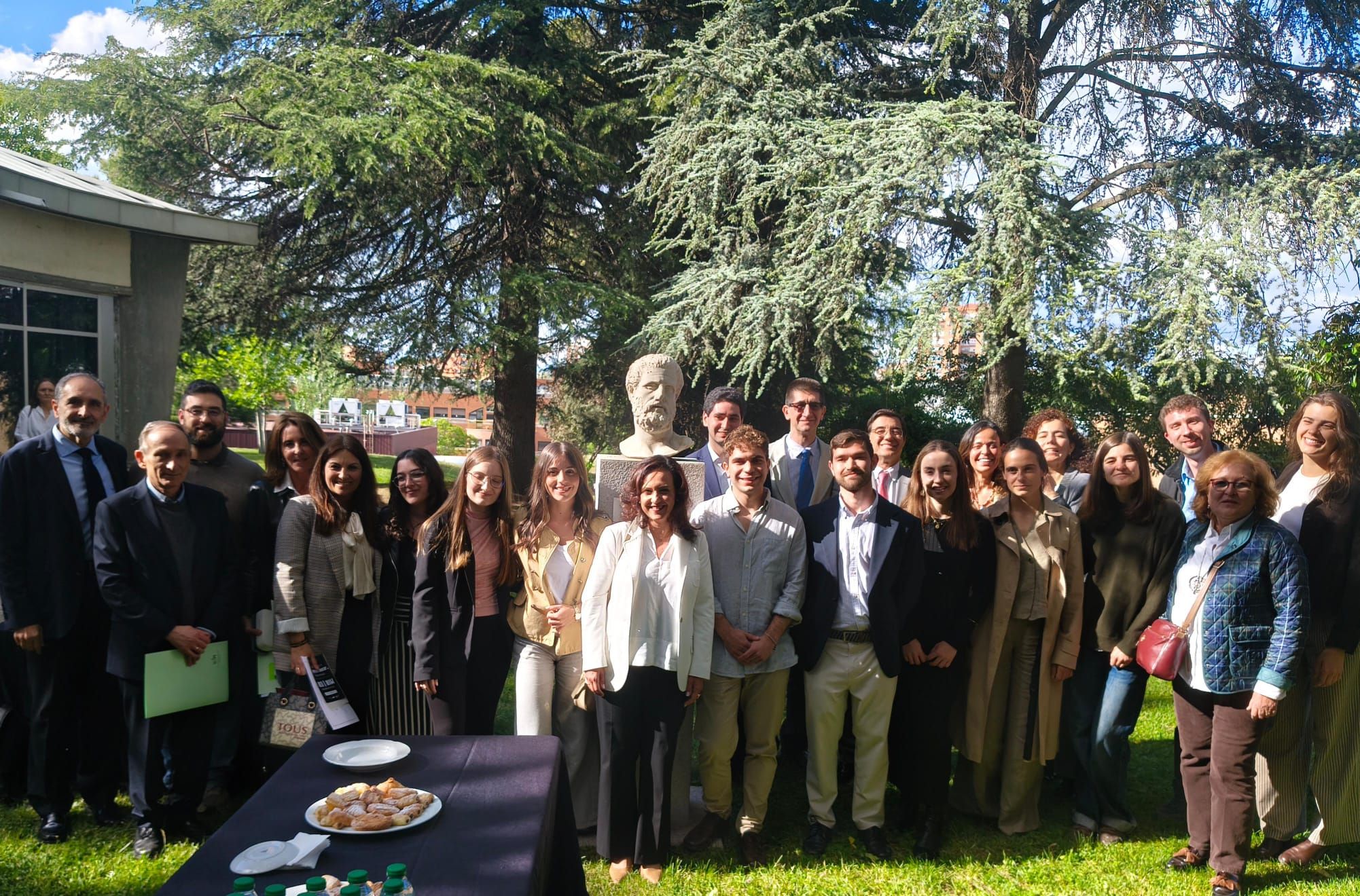
Celebrado el Acto de reconocimiento MIR/EIR y la primera edición de los «Premios Jesús Saldaña» a Tutores Clínicos
Celebrado el Acto de reconocimiento MIR/EIR y la primera edición de los «Premios Jesús Saldaña» a Tutores Clínicos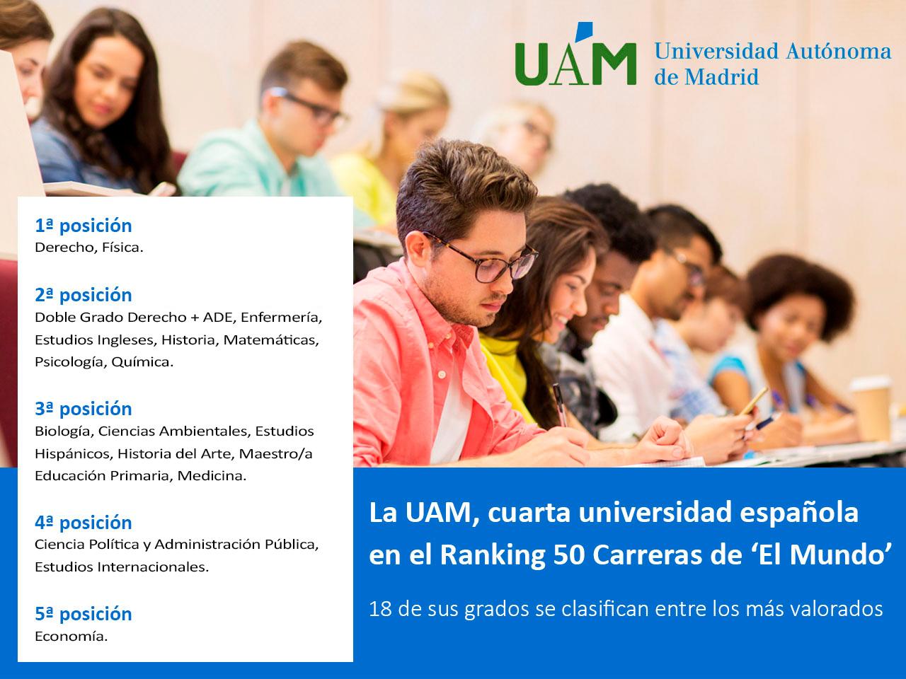
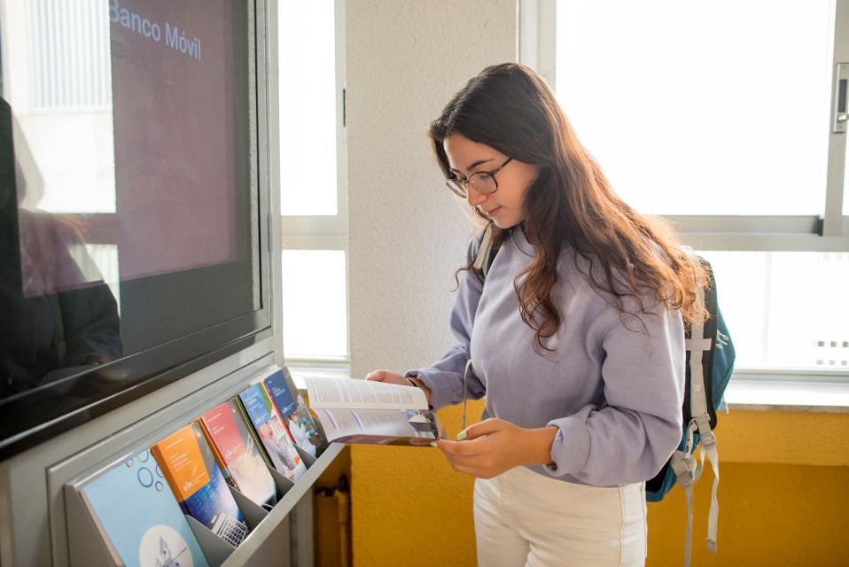
Enfermería y Medicina de la UAM destacan en el Ranking 50 Carreras de El Mundo 2026
Enfermería y Medicina de la UAM destacan en el Ranking 50 Carreras de El Mundo 2026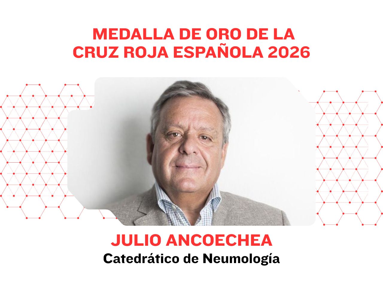
El doctor Julio Ancochea, Medalla de Oro 2026 de Cruz Roja Española
El doctor Julio Ancochea, Medalla de Oro 2026 de Cruz Roja Española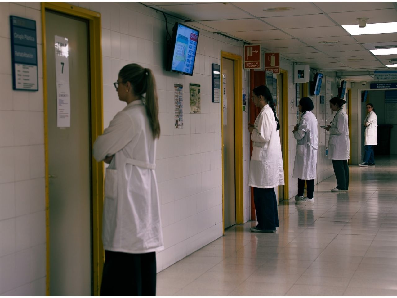
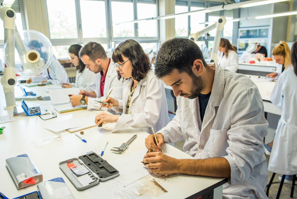
Celebrada la Prueba ECOE-UAM 2026 en el Hospital Universitario La Paz
Celebrada la Prueba ECOE-UAM 2026 en el Hospital Universitario La PazAgenda

Consulta la agenda de la Facultad.